Reconstructive Surgery
Facial Injury
Among general bodily traumas, head injuries are of particular importance because they lead to both functional and aesthetic losses.
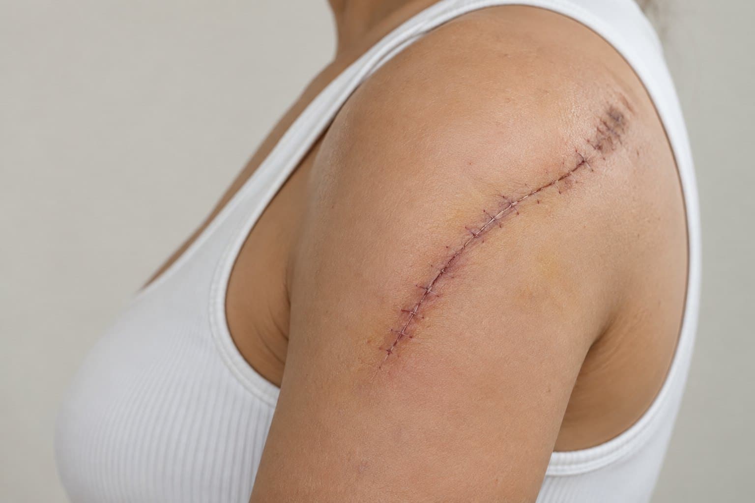
About Facial Injury
What is Facial Injury?
Among general bodily traumas, head injuries are of particular importance because they lead to both functional and aesthetic losses. In daily life, head and face injuries occur for many reasons, from simple household accidents to serious traffic accidents, and from sports injuries to injuries resulting from assault or fights.
Because of its complex three-dimensional anatomy [it contains almost all of our sense organs (sight, hearing, taste, smell), it contains the central nervous system (the brain, cerebellum and upper part of the spinal cord), and it contains the beginnings of the respiratory and digestive systems (such as the oral and nasal cavities and the pharynx)], the head region is an area that requires surgical interventions to be performed by skilled hands.
Although head and face injuries are sometimes isolated (on their own), ninety per cent of patients have another accompanying trauma. Loss of consciousness after the trauma is reported in 40% of patients. One third of patients have an accompanying limb injury or chest injury. It should not be forgotten that, unfortunately, 8% of patients with head and face injuries are lost. For this reason, after patients with head and face injuries are evaluated, interventions should be ordered according to medical importance. In life-threatening injuries, priority must be given to procedures that will save the patient's life; next, procedures that will result in permanent functional losses if delayed must be performed; and last, procedures that will reduce aesthetic losses. Although the ideal is to try to perform all surgical procedures at the same time as far as possible, sometimes this may not be possible for medical reasons.
In head and face injuries, bone fractures and soft tissue injuries (facial lacerations, facial nerve lacerations, and injuries of the salivary gland, salivary duct, tear duct, eyelid, lip, nose and ear) should be evaluated together. Stopping existing bleeding and securing the airway is life-saving for the patient. A neck collar must be applied to patients, and it should not be removed unless a neck X-ray is taken and it is confirmed that there is no fracture.
In the examination of patients with head and face injuries, painful areas in the soft tissue, areas with loss of sensation, double vision, facial asymmetry and dental closure problems need to be evaluated. In addition, visual acuity and examination of the inside of the mouth and nose must be assessed in every patient.
The usefulness of the plain X-rays recommended for evaluating bone structures is now debatable. A detailed computed tomography scan of patients saves both money and time. Thanks to computed tomography, the state of the brain and facial bones can be clearly shown, and if the procedures to be performed are considered necessary, they can be simulated in a computer environment to prepare for surgery. In addition, the three-dimensional images obtained provide an opportunity to give patients and their relatives clearer information about the planned operation(s).
Classification of facial fractures:
- Nasal Fractures
- Upper and Lower Jaw Fractures
- Jaw Joint Fractures
- Cheekbone (Zygomatic) Fractures
- Orbital Floor Fractures
- Frontal Sinus Fractures
- Complicated Fractures of All Facial Bones (Panfacial)
- Closed Methods Used in Facial Fractures
- Use of Distractors in Facial Fractures
Today, the gold standard in the treatment of facial fractures is to bring the fracture ends together with open surgery and fix the fractured pieces with tiny plates and screws. While titanium plate-screw systems are preferred in adult patients, self-absorbing (absorbable) systems are preferred in paediatric patients.
Although there are closed treatment options (without making an incision in the patient) in special situations such as isolated nasal fractures and cheekbone fractures, if doubt arises during the operation about the success of closed methods, switching to the open method will always be in the patient's interest.
In patients whose bone fixation has been performed correctly, the postoperative period is relatively painless. Patients are bothered by swelling and bruising rather than pain. Both complaints subside by themselves within a week to ten days after the operation. In patients in whom all the facial bones are fractured or the lower and upper jawbones are fractured, it may be necessary to fix the lower and upper jaws to each other (to perform double jaw fixation). These patients will need to be fed with soft, semi-solid foods for a period of 2-6 weeks. Patients with nasal fractures may have packing inside the nose and a plastic splint on the nose for about a week.
The metal plates and screws used for bone fixation are made of titanium, which is compatible with the human body. Titanium does not corrode in the tissues and is not harmful to the human body. Although they can be removed after the few months needed for the fractured bones to heal have passed, since this generally requires a second operation, they can be left on the bones without being removed. However, if they cause pain or cold intolerance in patients, or if they are superficial enough to be felt under the skin when touched (especially around the eyes), they are removed at the patient's request. Although titanium implants are MRI-compatible (they do not heat up and cause tissue damage during MRI), they impair image quality. For this reason, if patients who have had bone fixation with titanium plates and screws need to have an MRI for any reason and the image quality is not sufficient, the implants may need to be removed.
Let's Create the RightPlan for You Together.
Contact us for detailed information on Facial Injury and a treatment plan tailored to you.
First Step